

Background
AIG Hospitals in Hyderabad has used augmented and mixed reality headsets alongside a robotic exoscope to carry out a complex skull-to-spine tumour surgery, a procedure reported as a first for India's Telugu-speaking states.
Tumours that extend from the skull base down into the spine sit in a narrow, high-risk corridor crowded with nerves and blood vessels. Visualisation, rather than instrumentation, is often the limiting factor in operating on them.
The deployment
The operation was led by Dr Abhirama Chandra Gabbita, with Dr Subodh Raju, Director of Neurosurgery, part of the team describing the approach. The hospital combined two visualisation technologies in a single procedure rather than trialling them separately.
Mixed reality in the operating theatre
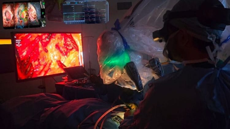
Wearing AR/MR headsets, the surgical team worked in an environment where digital anatomical maps and the physical operating field co-existed in real time. Instead of turning away from the patient to read a conventional monitor, the surgeons had 3D imaging rendered directly in their line of sight, keeping attention on the surgical field while referencing the underlying anatomy.
The robotic exoscope
Alongside the headsets, the team used a robotic exoscope as a second visual channel. Positioned over the operative site, it delivered magnified, high-clarity video of deep neural pathways within the high-risk corridor, giving the team a detailed view of structures that are difficult to reach and difficult to see.
Assessment
Dr Raju and Dr Abhirama characterised the combination as the direction operating theatres are heading. In their account, the technology removes the need to rely on guesswork or bulky screens: the headsets let surgeons look through tissue, while the robotic camera supplies a microscopic view of hard-to-reach spots. Both were explicit that the setup supplements rather than replaces the surgeon's hand, adding visual clarity rather than taking over any part of the procedure.

