

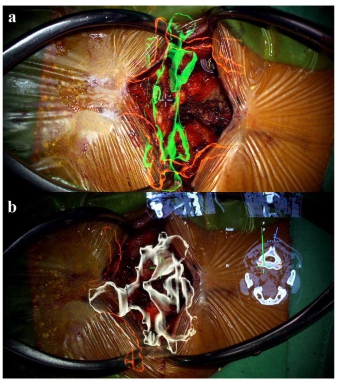
Taichung Veterans General Hospital reported microscope-based AR guidance for C1-C2 fixation in a patient with os odontoideum. The surgical team combined preoperative MRI and CT angiography with intraoperative CT, aligning colour-coded 3D bone and vertebral-artery models with the operative view. Tracking references on the skull clamp and microscope supported registration. Surgeons viewed planned screw entry points and trajectories through the KINEVO 900 microscope, integrated with Brainlab Curve navigation.
The May 2024 case report describes correct screw alignment, symptom relief and discharge on postoperative day four without neurological deficit. This is one clinical case, without comparative proof of improved accuracy or reduced operating time. Its operation date is not stated.
The hospital’s dedicated spine-surgery service page dates its wider microscope-based AR C1-C2 programme to 2021. A separate 2023 paper covers 67 patients treated across 2004–2022 using evolving techniques; that total is not an AR patient count.

