


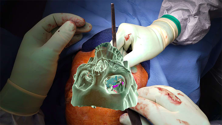
UC Davis Health's Medical Extended Reality (MXR) research group — surgeons from otolaryngology, neurological surgery and orthopaedic surgery working with the health system's 3D Printing and Visualization Lab — has taken augmented reality navigation into the operating room for craniomaxillofacial and head-and-neck surgery. The navigation platform was developed collaboratively with Xironetic, a surgical software company based in Oklahoma City. Between 20 September 2023 and 20 October 2024, 33 operations were performed with it at four tertiary teaching hospitals: UC Davis Medical Center in Sacramento (25 cases), David Grant USAF Medical Center at Travis Air Force Base in Fairfield, California (5 cases), University Hospital Freiburg in Germany (2 cases) and University Hospital Basel in Switzerland (1 case).
How it works: thin-slice CT (0.625 mm or finer) and MRI data are used to build a virtual surgical plan in Brainlab Elements and the open-source 3D Slicer, typically including mirrored anatomy from the unaffected side and any patient-specific implants. The plan is loaded into Xironetic's IntraOpVSP software, which runs entirely on a Microsoft HoloLens 2 headset and needs no external computer, camera tower or navigation cart. In theatre a reference array — either infrared reflective spheres or a printed ArUco optical code — is fixed to the patient's skull or, for most jaw cases, to a patient-specific dental splint. The surgeon touches a series of predefined anatomical landmarks with a tracked stylus, and the software locks the virtual anatomy onto the real patient, showing the overlay so registration accuracy can be checked immediately.
Once registered, the plan is visible heads-up on the patient rather than on a flat monitor across the room. Each anatomical object is named before surgery, and the surgeon calls it up by voice ("show zygoma", "hide zygoma") or by resting their gaze on a drop-down menu item for two seconds, so nothing has to be touched. An assistant dons and doffs the headset in under a minute to preserve sterility, and 2.5x to 3.5x surgical loupes fit underneath.
The 33 cases were 13 trauma, 8 orthognathic, 7 tumour and 5 craniofacial procedures, in patients from one month to 79 years old. Direct projection of the surgical plan was judged a significant advantage in all 33 cases; visualisation of hidden anatomy — tooth buds, the inferior alveolar nerve, dural sinuses, the optic canal, tumour margins — in 19 of 33; and real-time tracking of instruments and mobile bone segments in 10 of 33. The team reports several first-known uses: instrument tracking for midface osteotomies, tracking of the zygomaticomaxillary complex during fracture reduction, mandibular tracking in orthognathic surgery, augmented reality fibula cutting guides for mandibular reconstruction, and real-time infrared visualisation of vasculature through the headset, which showed a patient's external jugular vein that was not visible under white light alone.
The write-up is candid about the limits. Registration accuracy depends on careful landmark selection and can go wrong; virtual objects can drift, either apparently (as the surgeon moves around the patient) or genuinely, when the headset loses an accurate registration; opaque virtual objects can hide the very anatomy underneath them; and because the HoloLens 2 has no headlight attachment, high-intensity overhead lights have to be pulled away during 3D visualisation, which is awkward in deep cavities. Registration and utilisation times were not recorded, and the series is retrospective with no control group and no formal accuracy measurements. Three of the authors declare founding or co-founding roles at Xironetic and the senior author consults for Brainlab, so the clinical judgements are the operators' own.
Xironetic's IntraOpVSP software was cleared by the US Food and Drug Administration in October 2022 (510(k) K213128). Alongside the operating-room work, UC Davis Health uses the same extended reality toolkit to walk patients through their own scans before surgery and to teach residents and medical students, and the group's laboratory is due to move into the Aggie Square research campus on the health system's Sacramento site.

