


Background
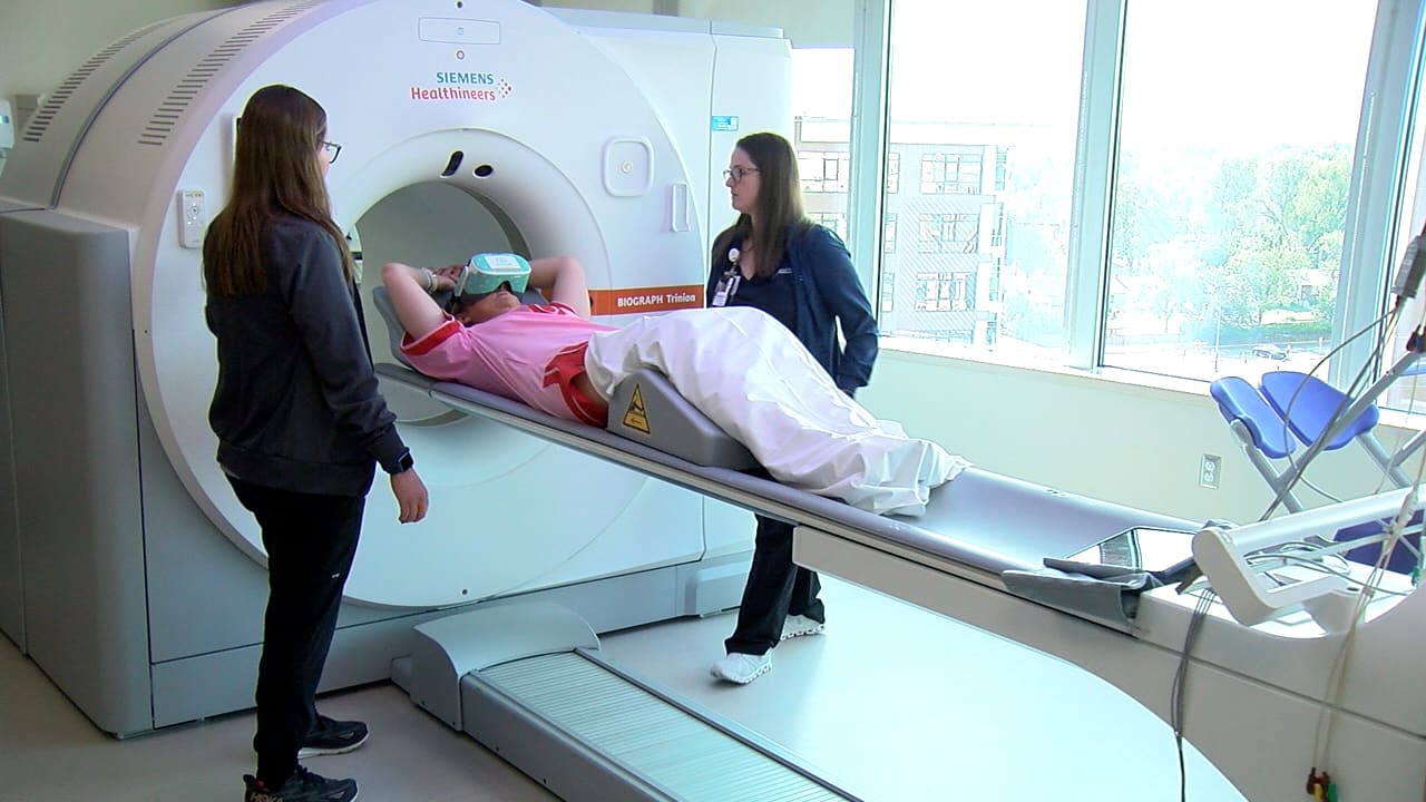
UCHealth Memorial Hospital Central in Colorado Springs uses cardiac CT to diagnose heart conditions including blocked arteries and infections. The exam requires the patient to lie inside the scanner's gantry, positioned from head to knees, and to stay still while the images are captured.
That enclosed position is difficult for patients who experience anxiety or claustrophobia. Stressed patients tend to move, and movement degrades image quality. A scan that cannot be completed has to be stopped or rebooked, which delays the diagnosis and the decisions other physicians make about that patient's care.
The deployment
The hospital's noninvasive cardiology team gives patients a virtual reality headset to wear during the scan. Instead of the inside of the gantry, the patient sees a calming environment; the options include hot air balloons, horse riding, ice skating, swimming with dolphins and relaxing on a beach.
Keeping the patient absorbed in the scene keeps them still, which lets the team capture the high-quality images the diagnosis depends on. Jamie Schmitt, manager of noninvasive cardiology at the hospital, says she has not come across another team applying the technique to cardiac CT.
Outcomes
Most patients have completed their procedure since the program began. Distraction has also carried patients through side effects that arise mid-scan: Schmitt describes a patient who started to feel nauseated, was distracted by a scene of penguins, and finished the exam.
The approach has drawn attention from the hospital's imaging supplier. Representatives from Siemens, on site for a follow-up about a new camera, saw the goggles in use and told the team they knew of nobody else doing it.
What's next
The cardiology team is working on ways to tailor the experience to the individual patient. Schmitt said future versions may let patients supply their own photographs or music videos to be uploaded and played during the procedure.


